Rhizarthrosis: Thumb basal joint osteoarthritis
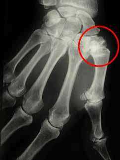
Thumb basal joint osteoarthritis is an arthrosis affecting the base of the thumb which is the progressively destroy the joint cartilage between the trapezius and first metacarpal.
Diagnosis

Arthrosis affecting the base of the thumb, i.e. the progressive destruction of the joint cartilage between the trapezius and first metacarpal. This occurs frequently especially in women and generally begins around age 50. It can also be the result of a knock or fracture.
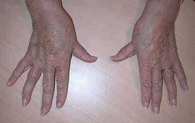
This arthrosis can be well-tolerated in certain cases or else lead to pain that constitutes a handicap to everyday activities and is associated with a Z-shaped deforming of the thumb bone.
Diagnosis is made on examination and by questioning the patient. Pain is observed on pressure to the base of the thumb or during manipulation. The inability to open the first commissure, a Z-shaped malformation with hyper-extension of the joint between the first metacarpal and first phalanx may also be seen.
X-ray assessment is usually requested.
Treatment

Initially, treatment is of a medical nature with analgesic and anti inflammatory medication. A resting splint may be worn at night.
One or several corticoid infiltrations to the joint can be prescribed to calm the pain.
When medical treatment fails, surgery may be proposed.
Several types of procedure are possible, with ablation of the trapezious and stabilisation of the thumb by means of a tendon in the vicinity, the complete blocking of the joint, or the placing of a prosthesis.
Personally I tend to use the technique consisting in removing the trapezious and stabilising the base of the thumb using a tendon in the vicinity.
For early stage painful arthrosis, especially for active people, a mini-invasive arthroscopic procedure can be proposed, with short time immobilisation and early recovery of the function.
Surgical procedure

- Length of hospital stay: two to three days
- Anaesthetic: regional (only the arm is put to sleep)
I most often propose a trapezectomy – ligamentoplasty.
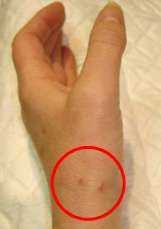
An incision is made on the outer edge of the wrist.. The worn down trapezius bone is completely removed. Part of the long protractor ligament of the thumb situated directly next to it is taken to stabilise the base of the thumb and serve as a « shock absorber ». Binding is sometimes realised and is left in place during the immobilisation period in order to stabilise the structure.
After-surgery care
- Immobilisation will last for a total of six weeks using a splint, initially in resin and then replaced by a made-to-measure plastic splint which includes the base of the thumb and the wrist but leaves the long fingers free.
- The fingers may be used normally under protection of the splint.
- Physical therapy is commenced after the immobilisation period and is necessary for the recovery of full range of movement.
- Even if it is possible for the patient to recover the use of his hand progressively after a month and a half, the final result is only appreciated after approximately six months. By that time pain has clearly improved and mobility has been well preserved.
Possible complications
Local infection is rare but remains possible : a swollen, painful and stiff hand may be seen in cases of Algoneurodystrophy which may develop over several months. It is not possible to predict its occurrence or the stiffness left as an after effect and pain is therefore a possibility Pain spanning over the thumb or shooting up to the wrist may persist over several months and is linked to the regional tendinitis.
In some patients, recovering mobility can prove difficult and pain may persist.
Tingling sensations in the hand may be observed before or after such a procedure and are linked to carpal canal syndrome. This phenomenon is not caused by surgery, but often associated with the initial arthrosis itself.